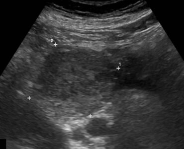
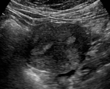
Complicated Appendicitis

Acute Complicated Appendicitis in a 31-year Old Male

Patient History

A 31-year old presented with 1 day history of central abdominal pain radiating to the right iliac fossa. Raised WCC and  CRP.

Case Description

The patient was referred to have an ultrasound of the abdomen and pelvis to include the appendix. Ultrasound revealed an abnormal appendix in the RIF measuring 14 mm in AP calibre with significant probe tenderness observed while scanning the area. There was also evidence of mesenteric fat stranding around the inflamed appendix with hypervascularity within the appendix wall. An echogenic focus was seen within the appendix suggestive of a small appendicolith. 

Treatment/ Follow Up

The patient had laparoscopic appendectomy and the appendix specimen was sent for histology analysis.

Histology revealed an inflamed appendix with focal mucosal ulceration in keeping with complicated appendicitis.

Leave a Comment

Share this case

Acute Complicated Appendicitis in a 31-year Old Male

Or copy link

Table