• Home
  • Videos
    • How To’s
  • Patient Arena
  • Sonohive cases
  • Forums
  • Blog
  • Events
  • More
    • Resources
    • About Us
    • FAQs
Account
  • Sign In
  • Sign Up
Submit Case
Sonohive Sonohive
  • Home
  • Videos
    • How To’s
  • Patient Arena
  • Sonohive cases
  • Forums
  • Blog
  • Events
  • More
    • Resources
    • About Us
    • FAQs
Sonohive Sonohive
Sonohive Sonohive
  • Home
  • Videos
    • How To’s
  • Patient Arena
  • Sonohive cases
  • Forums
  • Blog
  • Events
  • More
    • Resources
    • About Us
    • FAQs
  • Sign In
  • Sign Up
Submit Case
Updated on December 16, 2024

Duodenal Tumour:  An Obstructive Duodenal Tumour in a 76 Year Old Patient, Presenting as Epigastric Pain and Haematemesis

295 Views
Share

Clinical History

A 76-year old man presented with epigastric pain and haematemesis discovered to be a duodenal tumour.

Case Description

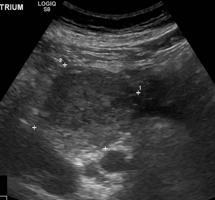
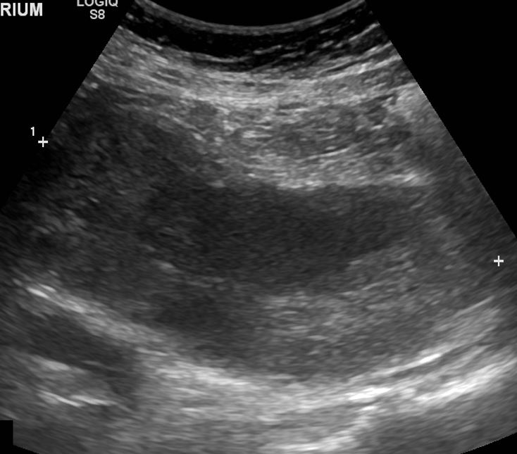
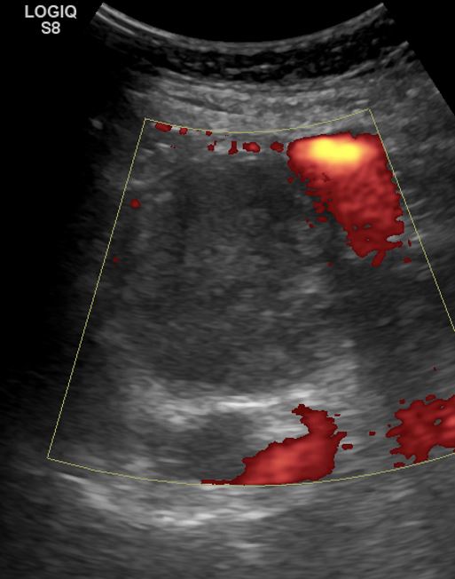
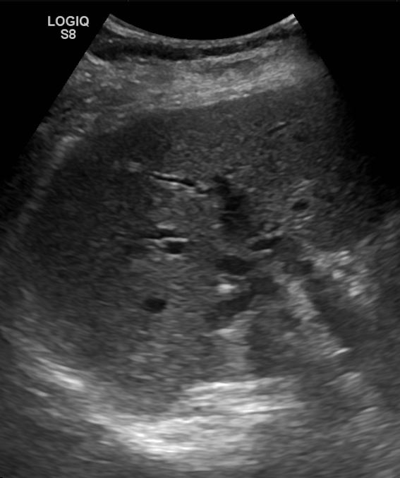
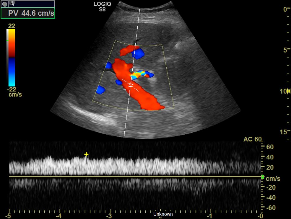
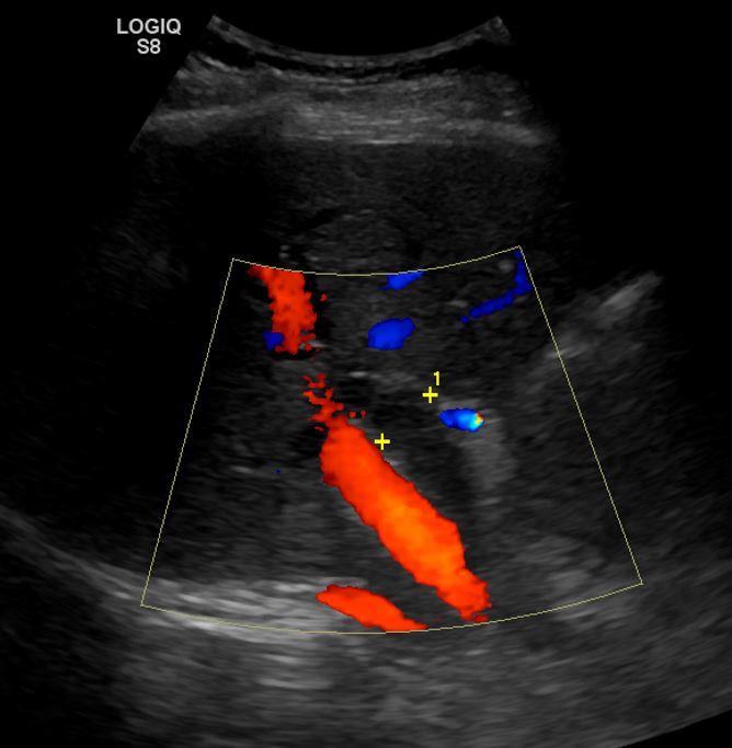
The patient was referred to have an abdominal ultrasound which reveals a large heterogeneous mass in the duodenum. There was also intrahepatic biliary dilatation and raised portal vein flow velocity, all secondary to the obstructive nature of the duodenal mass.

Treatment/Follow Up

The patient also had Oesophagoduodenoscopy (OGD) which revealed the mass to be causing a gastric outflow obstruction. 

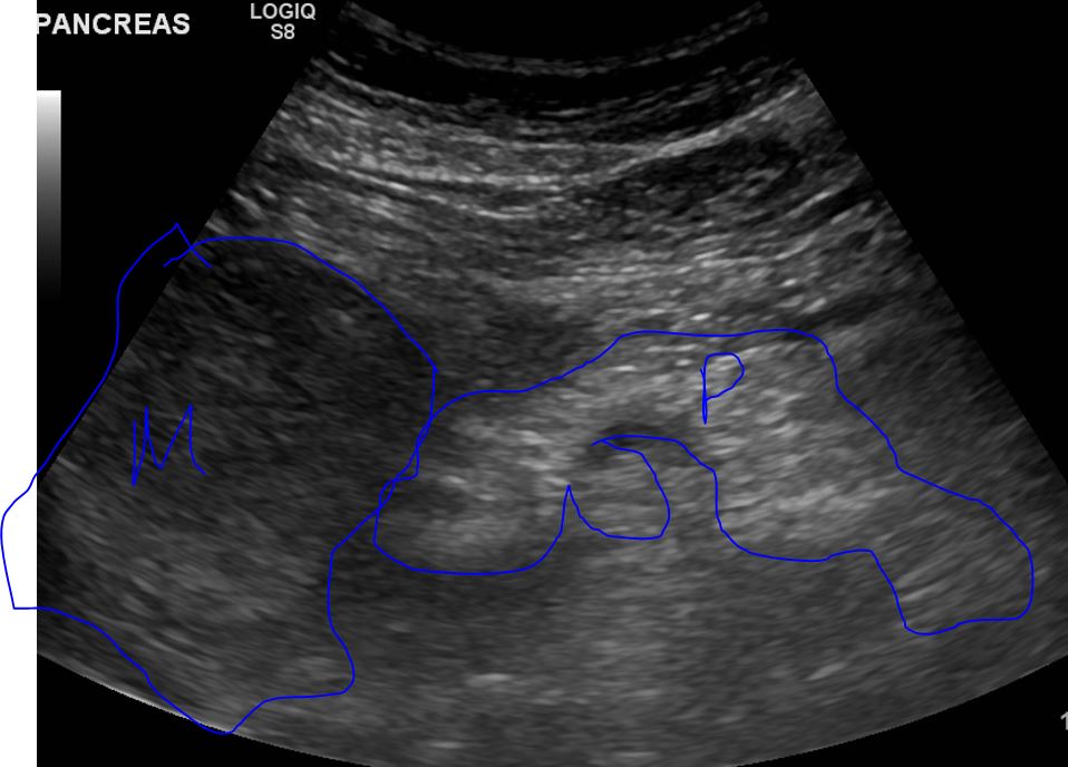
The patient also has a CT scan of the chest abdomen and pelvis (with contrast) which revealed the obstructive mass to be at D2/3 with an abnormal D3 and an abrupt calibre of D4. 

The patient was referred to the Upper GI specialists for further management.

Sonogram of the abdomen at the level of the epigastrium, showing the duodenal tumour
Sonogram of the abdomen at the level of the epigastrium, showing the duodenal mass
Sonogram at the level of the epigastrium showing the long segment of the duodenal tumour
Sonogram at the level of the epigastrium showing the long segment of the duodenal mass
Power Doppler imaging of the duodenal mass. No internal vascularity seen. However, there is evidence of peripheral vascularity
Sonogram of the pancreas (P) with the duodenal mass (M) seen adjacent and medial to the pancreatic head
Right hepatic lobe showing evidence of intrahepatic biliary dilatation secondary to the obstructive duodenal mass
 Patent portal vein with a slightly elevated velocity measurement of 44.6 cm/s due to the duodenal mass-effect on the proximal portal vein
 Patent portal vein with a slightly elevated velocity measurement of 44.6 cm/s due to the duodenal mass-effect on the proximal portal vein
Dilated common bile duct (CBD) secondary to the obstructive duodenal mass
Sonohive
Author: Sonohive

Tags : Abdominal scan Duodenal Tumour Epigastric Pain Haematemesis Small Intestine Sonogram Ultrasound

Leave a Comment Cancel reply

You must be logged in to post a comment.

Welcome to sonohive website, we feature comprehensive cases, patient arena, and a discussion forum. Connect, explore, and learn with us.
Facebook X-twitter Instagram Linkedin Youtube
Quick Acess
  • About Us
  • FAQs
  • Contact Sonohive
  • Resources
  • About Us
  • FAQs
  • Contact Sonohive
  • Resources
Legal
  • Privacy Policy
  • Terms of Use
  • Acceptable Use Policy
  • Authors Guide and Terms for submission
  • Privacy Policy
  • Terms of Use
  • Acceptable Use Policy
  • Authors Guide and Terms for submission
© 2024 All Rights Reserved by Sonohive Ltd

Duodenal Tumour:  An Obstructive Duodenal Tumour in a 76 Year Old Patient, Presenting as Epigastric Pain and Haematemesis

Or copy link

Clipboard Icon