Normal Doppler Ultrasound Assessment of a Transplant Liver
Clinical History
A 45-year old with a history of chronic polycystic liver and kidney disease had a recent liver transplant. Doppler ultrasound was requested to assess the blood flow in and out of the transplant liver.
Case Description
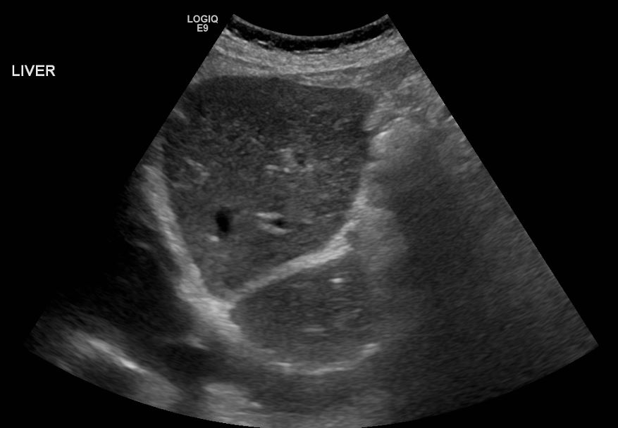
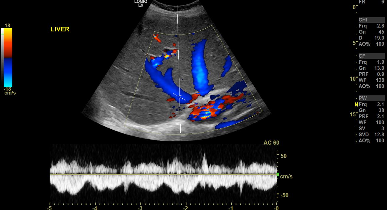
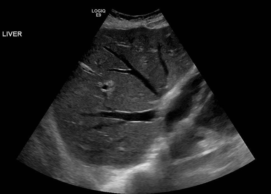
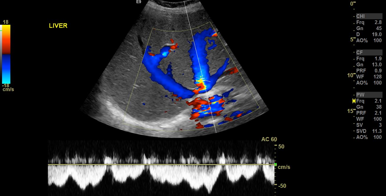
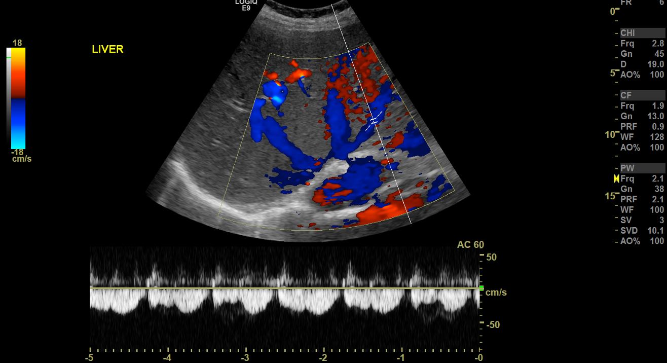
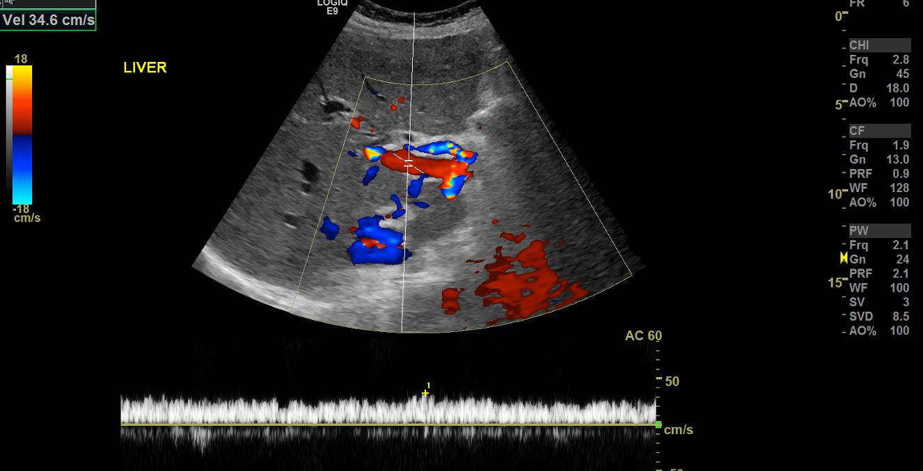
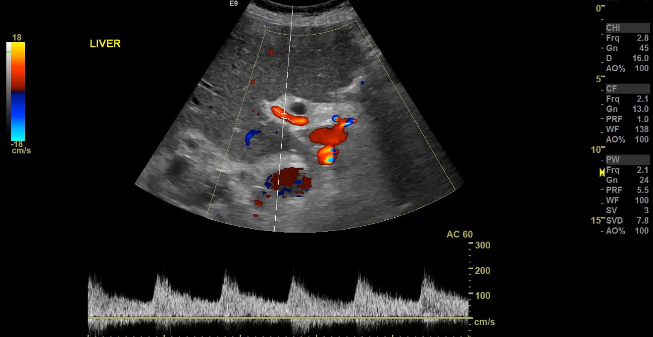
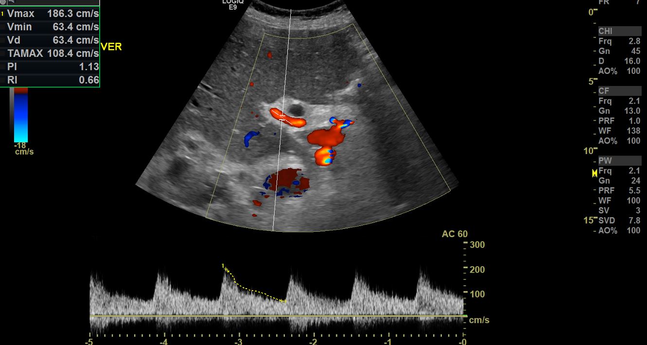
Ultrasound was performed using a 3 MHz curvilinear transducer. The examination started on B-mode to visualise the entire hepatic outline and parenchyma. Colour Doppler was used to evaluate patency of the hepatic veins, common hepatic artery, and the main portal veins and its branches. Spectral Doppler was further used to examine the flow pattern and velocity in the hepatic vessels.
Diagnosis/ Discussion/ Treatment/ Follow-up
The vessels examined in transplant liver assessment include:
The right hepatic vein.
The middle hepatic veins.
The left hepatic vein.
The main portal vein.
The right portal vein.
Middle portal vein.
Left portal vein.
The common hepatic artery.
The parameters deduced from the haemodynamic spectral Doppler studies include: peak systolic velocity (PSV), end diastolic velocity (EDV), pulsatility index (PI), resistivity index (RI), and the wave pattern.
Sonograms