Right Orchitis in a 30-year Old Male with a Coexisting Left Varicocoele
Patient History
A 30-year old male presented with an acute onset of right testicular pain.
Case Description
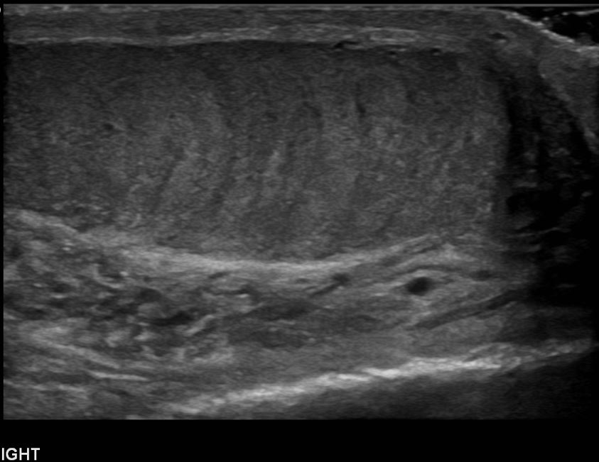
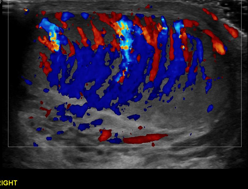
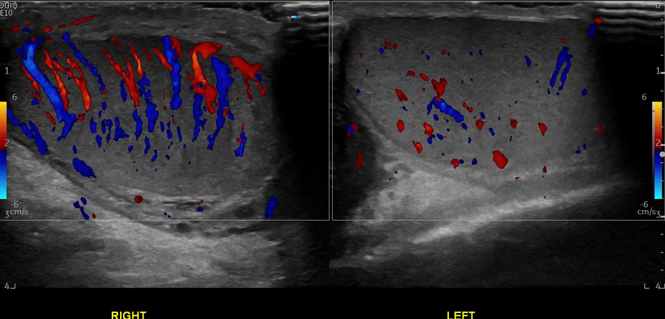
An ultrasound of the testes was performed using a 15 MHz linear transducer. Ultrasound revealed a hypoechoic and striated right testicle with evidence of hypervascularity on colour Doppler imaging. Appearances were in keeping with right orchitis.
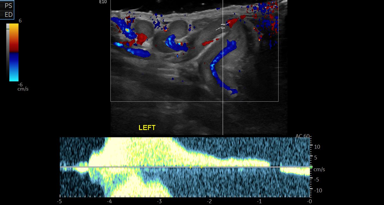
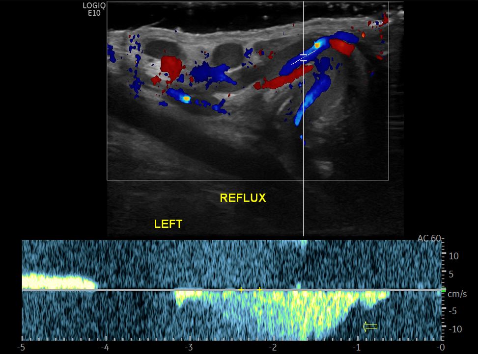
There was also evidence of dilatation of the left pampiniform plexus with a flow reversal of more than 2 seconds on spectral Doppler imaging. Appearances were suggestive of left varicocoele.
Diagnosis/ Discussion/ Treatment/ Follow up
The patient’s symptoms resolved after antibiotic therapy.
Sonograms