Urolithiasis in the Bladder and Kidney of a 75-year Old Man
Patient History
A 75-year old man presented with macroscopic haematuria
Case Description
The patient was referred to have an ultrasound examination of his kidneys and bladder as part of the (NICE guideline) diagnostic workup for haematuria in individuals above 45-years old.
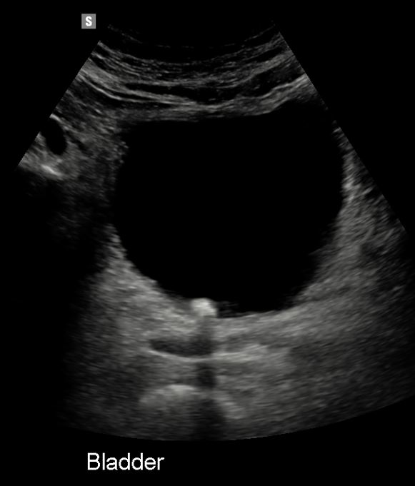
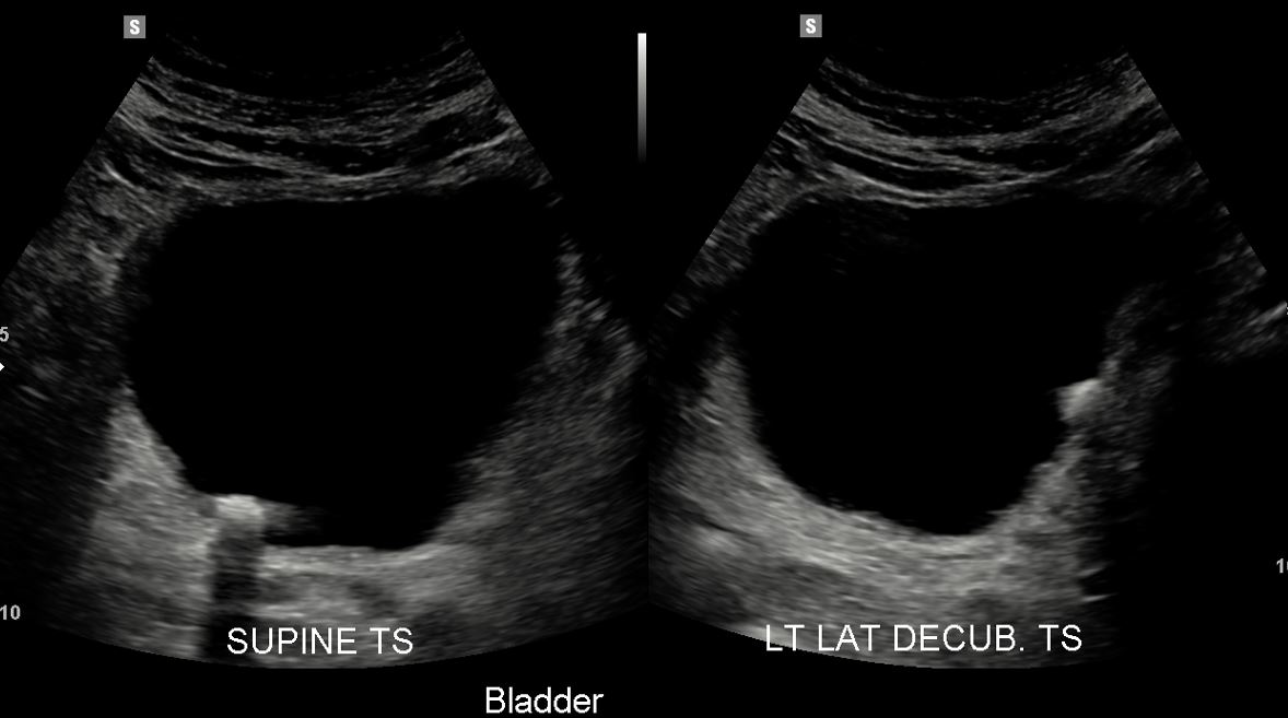
The bladder contained a 13 mm intraluminal mobile calculus (Urolithiasis).
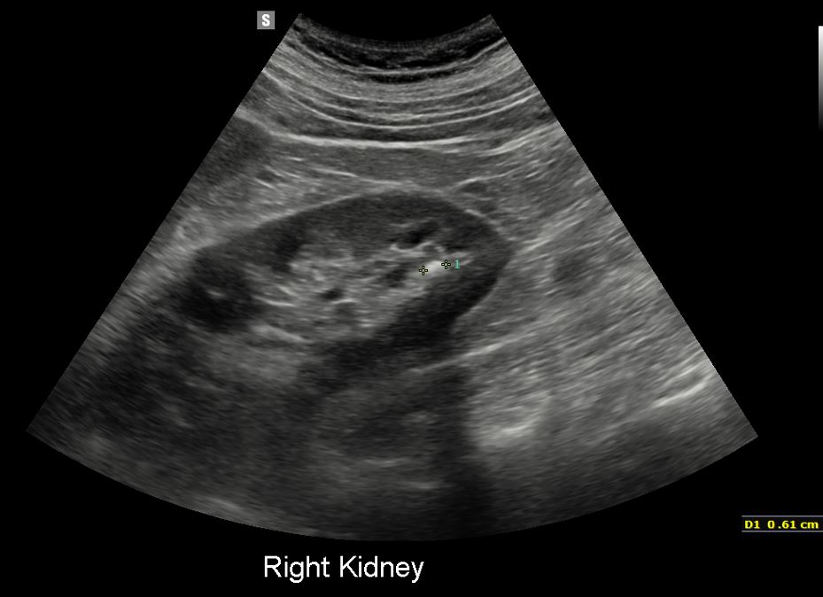
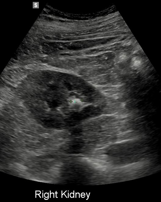
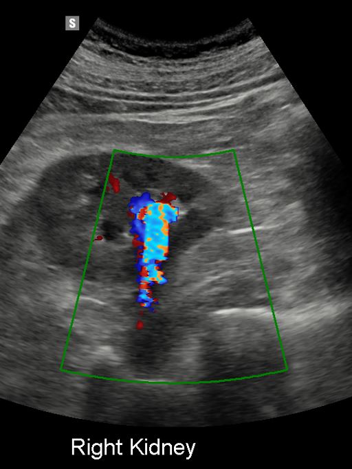
The right kidney contained an 8 mm non-obstructing calculus within its lower pole.
Diagnosis/ Discussion/ Treatment/ Follow Up
The patient had a follow up CT scan which confirmed the findings. The bladder calculus was removed transurethral.
Sonograms